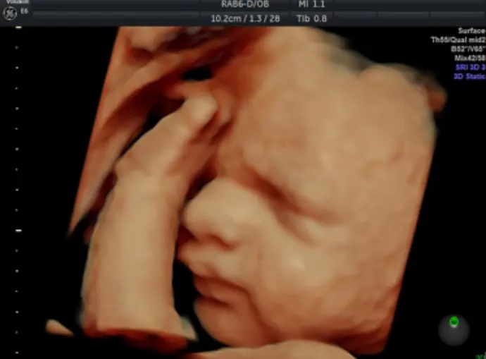
It is very common for expectant mothers to have a growth scan at around 32 weeks gestation, in order to make sure the baby is developing as normal.
In this blog post we'll answer some common questions about the 32-week growth scan, such as why it is needed, what happens during the scan, and what can the results tell you.
Why have a growth scan at 32 weeks?
The main goal of the 32-week pregnancy scan is to check that your baby is developing and growing as expected. Hospitals may recommend a scan at 32 weeks if they suspect too much amniotic fluid, or you can book a scan for your own reassurance.
The results of your growth scan will tell you:
- The position of your baby in the womb
- If your baby is smaller or larger than expected
What to expect from your 32-week growth scan
Your growth scan will be conducted in the exact same way as all your other pregnancy scans, with gel on your stomach and an ultrasound transducer device that allows you to see an image of your baby.
Our trained and qualified sonographer will be able to...
- Evaluate the volume of amniotic fluid surrounding your baby
- Measure your baby's head, abdomen and femur
- Measure the blood flow around your baby's body
What does the 32-week growth scan tell you?
The 32-week growth scan looks at your baby's wellbeing by measuring their biophysical profile. During the scan, the sonographer will be looking to see if your baby:
- Opens and closes their hands
- Stretches and flexes
- Moves their arms and legs frequently
- Makes breathing movements
During your growth scan, the sonographer will be able to see if your baby is smaller than expected for their gestational age, possibly due to a lack of nutrition through the placenta or restricted levels of oxygen.
The 32-week scan can also reveal if your baby is larger than expected. While this is usually not a medical concern, you may need to get tested for gestational diabetes if your unborn child is significantly larger than normal. This involves measuring the levels of glucose in your blood - high levels could result in birthing complications and enhance the likelihood of induced labour or a caesarean.
In regards to the position of your baby, your 32-week growth scan will show whether the baby is head down (normal position), feet first (breech position) or laying sideways (transverse position). Your doctor may advise you to have an ECV (external cephalic version) if your baby is in the breech position. This is a completely safe procedure whereby a surgeon pushes down and around on your abdomen in order to turn your baby into a normal head-down position for birth.
Book your 32-week growth scan now!
The following growth scans are available at 32 weeks:
- 4DGrowth&Wellbeing™
2D growth, reassurance and wellbeing scan with 4D bonding experience (24 - 32 weeks)
All of these scans are delivered at the First Encounters Ultrasound clinic in Cardiff. You may invite up to four guests to join you in the examination room, with five more guests in a separate viewing room (nine guests total, not including the mum-to-be).
If you have specific questions regarding any of our ultrasound scan packages, please do not hesitate to get in touch. Our friendly team members are always happy to help.
Browse Scan Packages
